
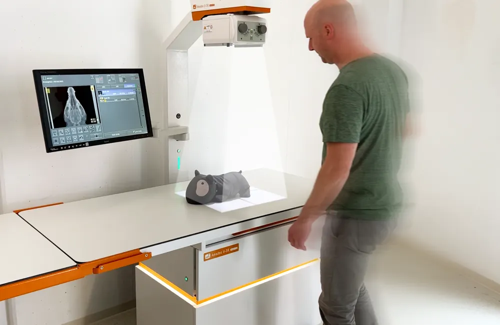
Digital premium complete X-ray solution for the modern small animal practice

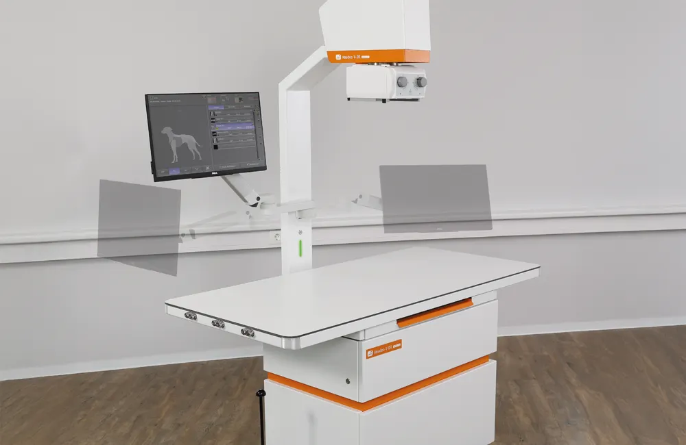
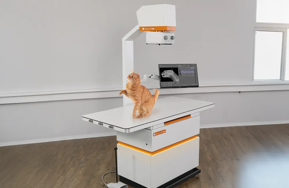
Amadeo V X-ray systems
Complete X-ray solutions for small animal practices with outstanding features
 Advice wanted
Our Amadeo V models
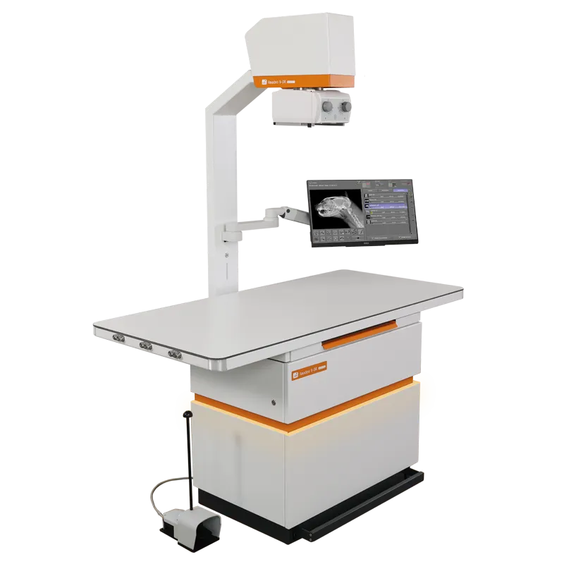
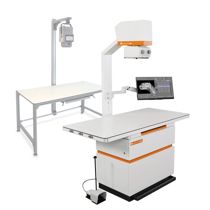
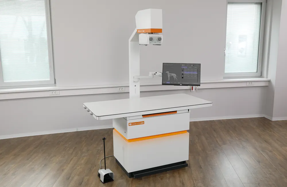
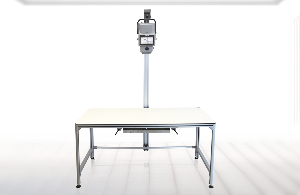
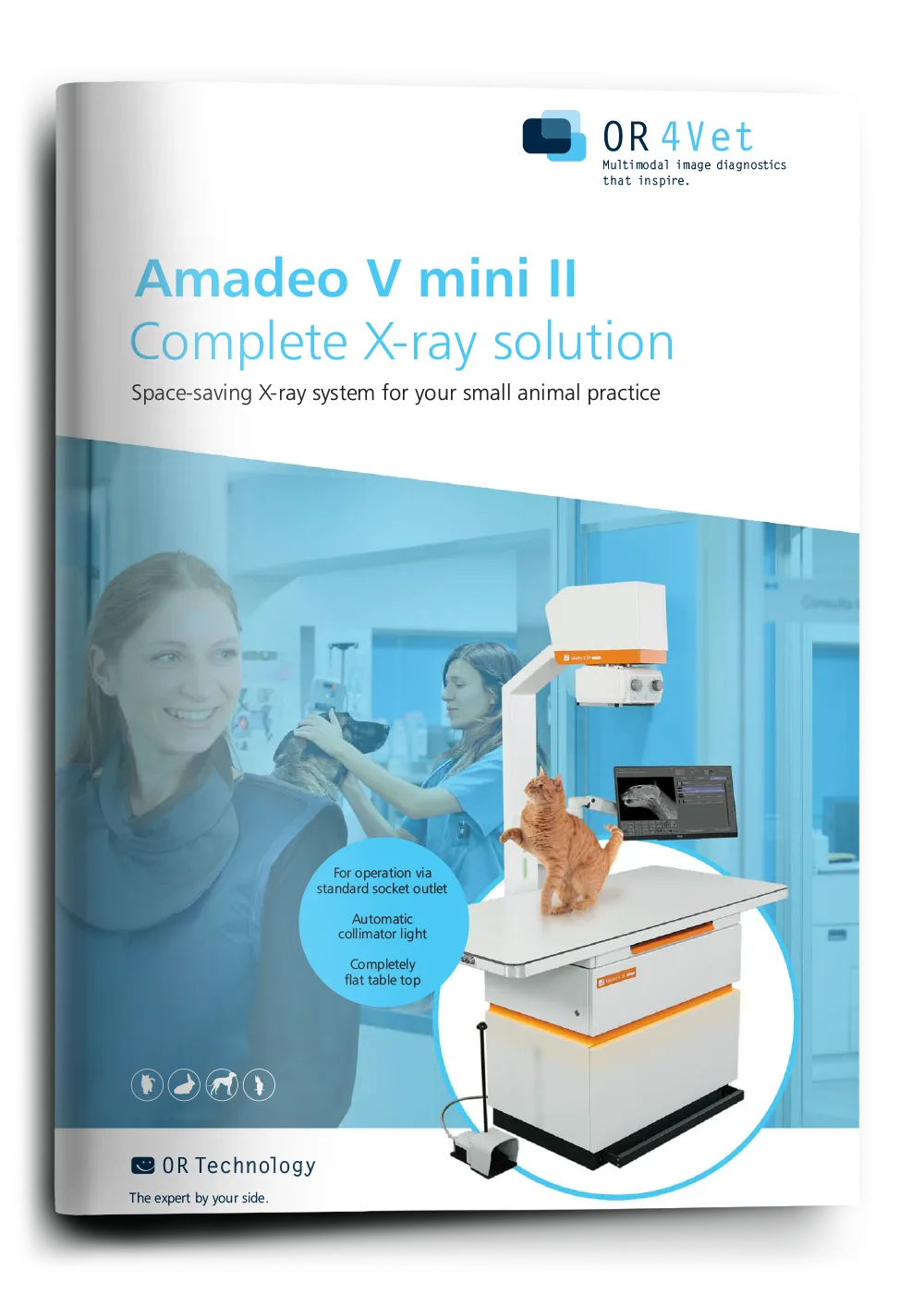
Amadeo V mini IITM
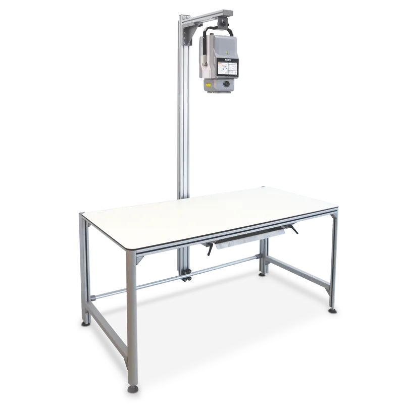
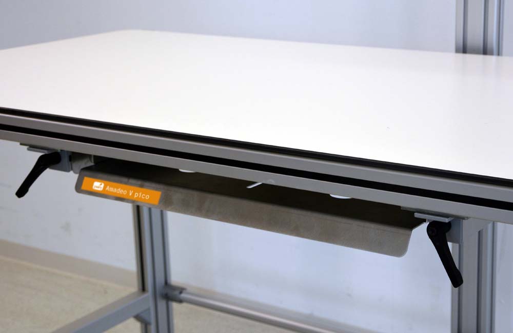
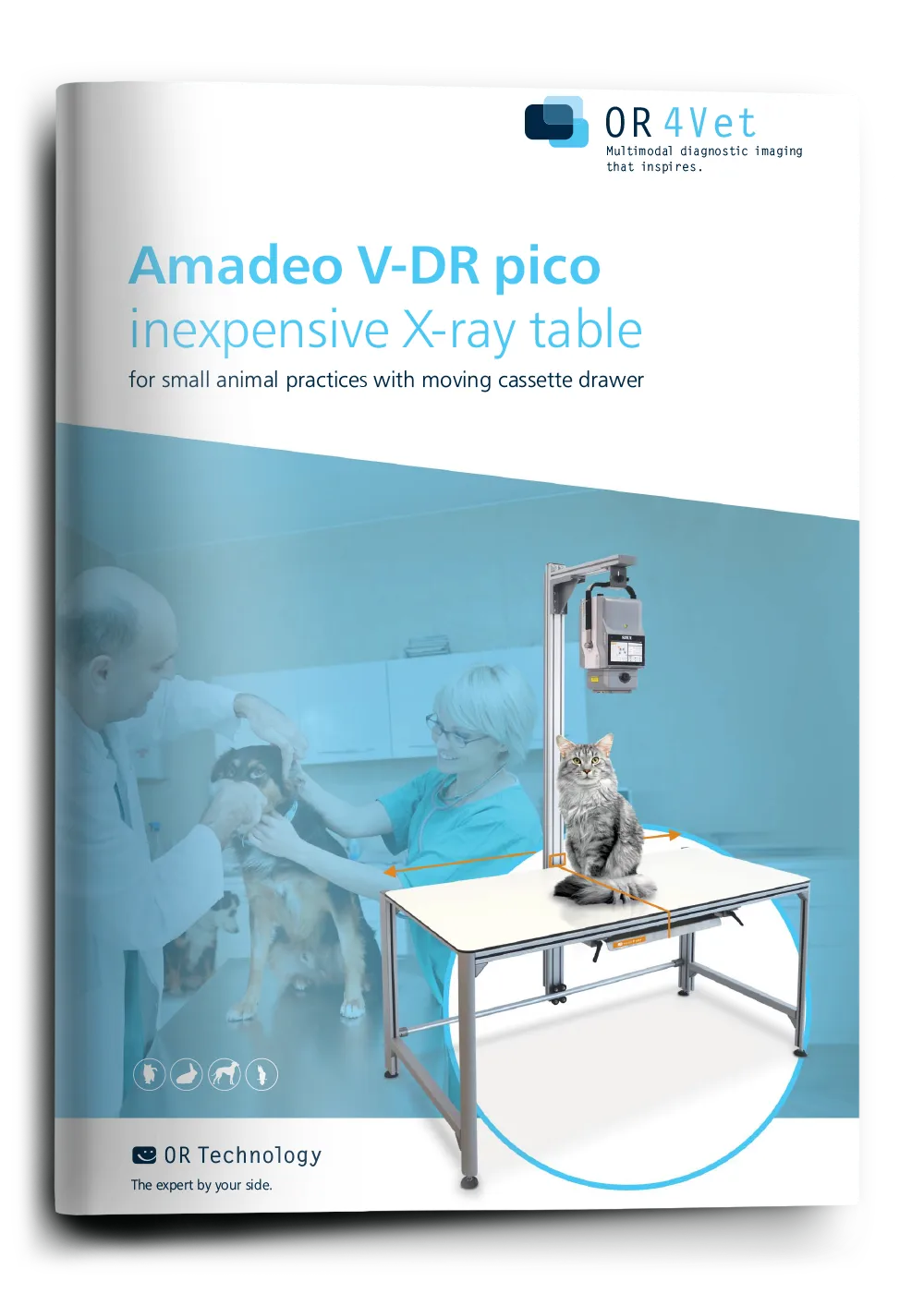
Amadeo V picoTM
Product Type
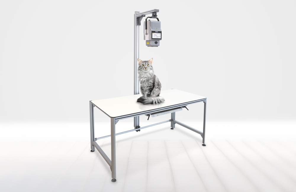
Compact, cost-effective X-ray table for flexible practice use
Applications & Benefits
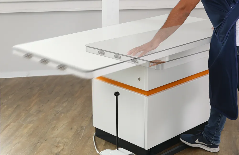
Ideal for fully digital X-ray diagnostics with maximum comfort, space-saving design, swivelling monitor, automatic collimator light
Perfect for cost-conscious practices – simple over-table and under-table exposures with integrated cassette drawer
System Performance
Powerful 32 kW HF generator, 230 VAC standard power socket
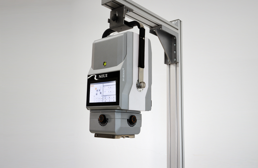
5 kW generator with large touchscreen, 230 VAC standard power socket
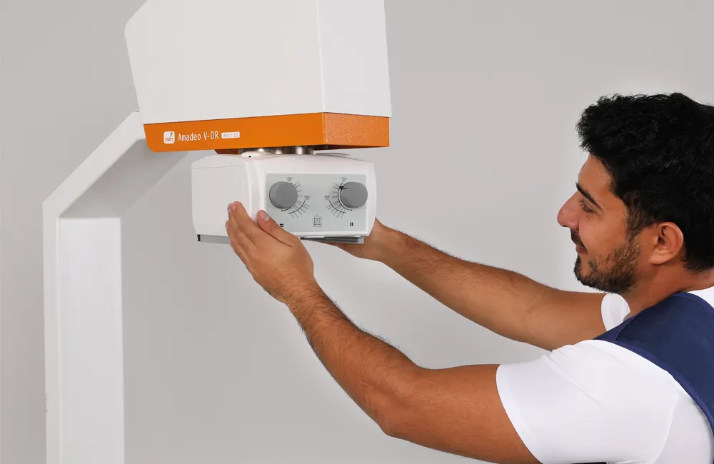
Operating Comfort
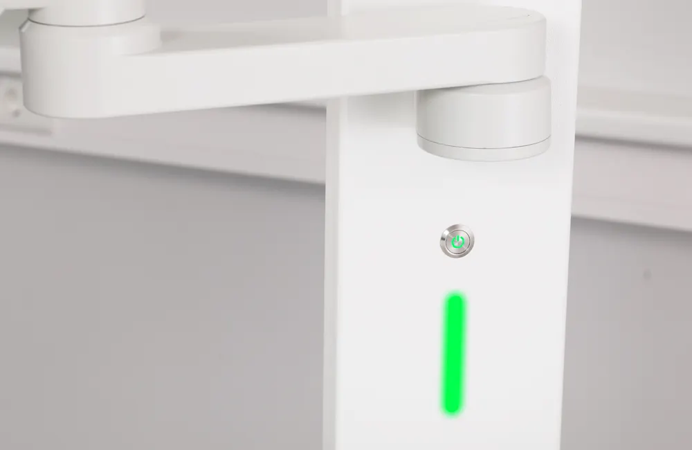
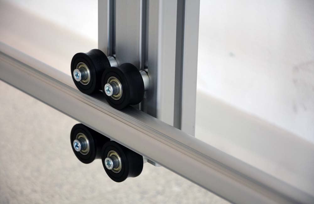
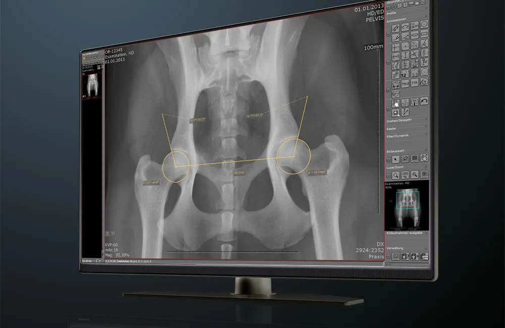
24″ touchscreen, integrated X-ray software, AI-supported measurement functions, 4-way floating table, 3-colour workflow visualisation
Easy positioning, robust and low-maintenance design, optional intuitive X-ray software
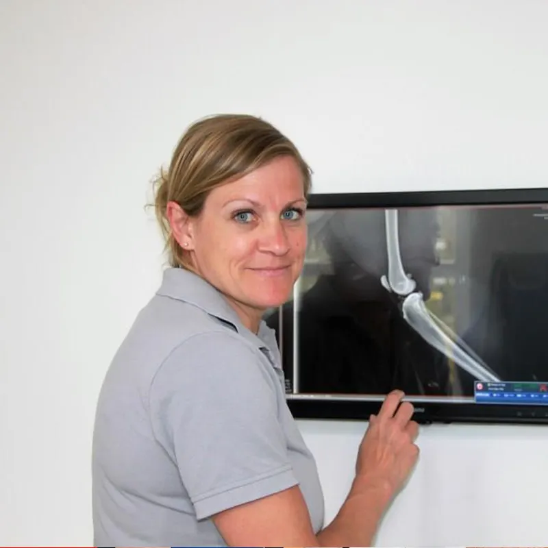
Image Quality
Excellent CsI detector – high level of detail at low dose
Flexible detector choice – solid image quality depending on system used
 Discover all details (PDF)
Amadeo V mini IITM
Product Type
Digital premium complete X-ray solution for the modern small animal practice
Applications & Benefits
Ideal for fully digital X-ray diagnostics with maximum comfort, space-saving design, swivelling monitor, automatic collimator light
System Performance
Powerful 32 kW HF generator, 230 VAC standard power socket
Operating Comfort
24″ touchscreen, integrated X-ray software, AI-supported measurement functions, 4-way floating table, 3-colour workflow visualisation
Image Quality
Excellent CsI detector – high level of detail at low dose
Amadeo V picoTM
Product Type
Compact, cost-effective X-ray table for flexible practice use
Applications & Benefits
Perfect for cost-conscious practices – simple over-table and under-table exposures with integrated cassette drawer
System Performance
5 kW generator with large touchscreen, 230 VAC standard power socket
Operating Comfort
Easy positioning, robust and low-maintenance design, optional intuitive X-ray software
Image Quality
Flexible detector choice – solid image quality depending on system used
Comprehensive and regional service
for your digital X-ray system
Our regional service team provides rapid support, maintenance and customised solutions – on site or via remote maintenance. This ensures that your X-ray system remains reliably operational.
Overview of services
Why buying from OR Technology
is the best decision for you
You can rely on the high quality of our products.
Thanks to international quality standards, you are relying on a safe and high-performance solution.
We offer you a customised software solution “Made in Germany”.
Rapid on-site assistance and comprehensive support by phone or remote maintenance are available to you at any time.
With thousands of systems installed worldwide, you rely on proven products trusted by many.
You can trust us thanks to our decades of experience.
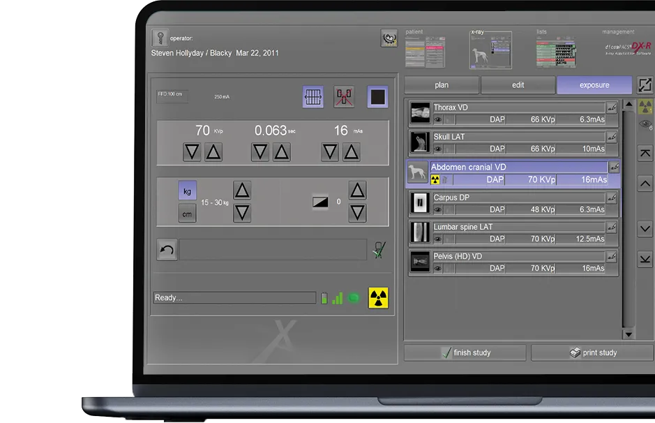
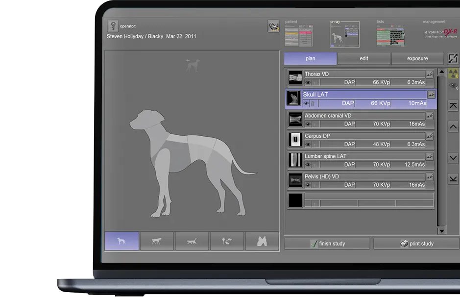
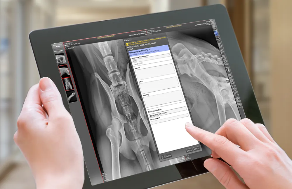
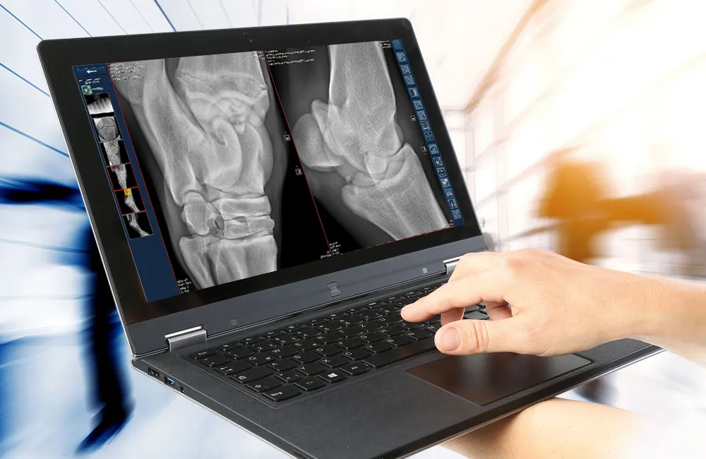
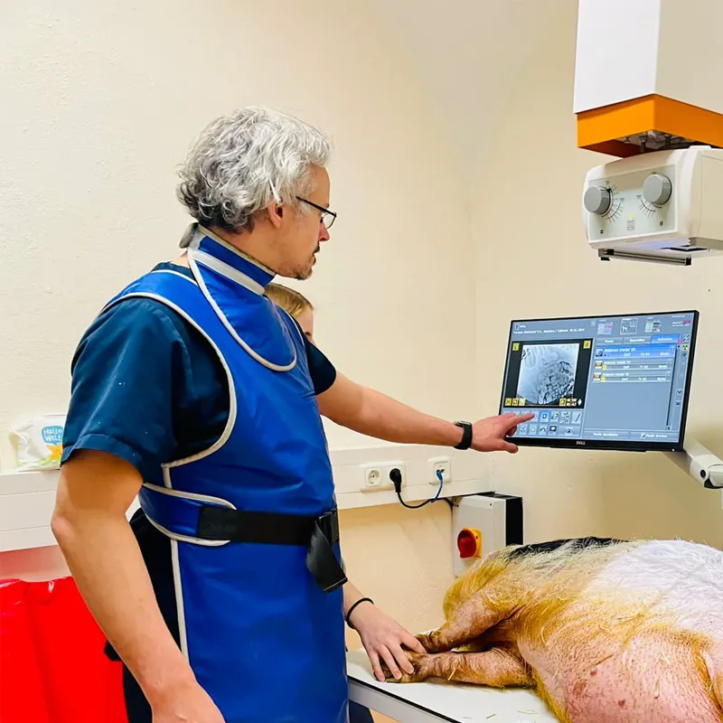
X-ray software that convinces…
dicomPACS®DX-R is the heart of our OR 4Vet systems – featuring smart operation, AI support, dynamic X-ray, and wireless control – perfectly tailored to modern veterinary medicine. A fast and reliable acquisition and diagnostic software that optimally supports you in everyday practice.
Discover X-ray solution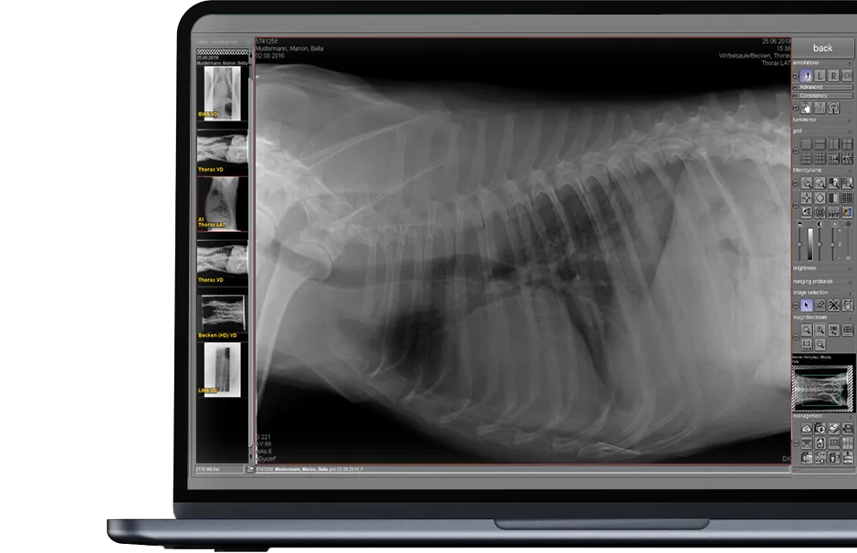
The full picture – in our catalogues
Product info
X-ray system Amadeo V mini II
Space-saving full X-ray system with rotating monitor arm for use with standard wall sockets
Product info
X-ray system Amadeo V pico
Minimalist and very cost-effective X-ray table including 5 kW generator and tracking detector loader
Product catalogue
PACS | X-ray | Ultrasound | CT
The guide for veterinary clinics and practices
Optional components
dicomPACS®MobileView
dicomPACS®MobileView is a web-based system that enables location-independent viewing and editing of radiological image data on mobile devices
Learn more (PDF)ORCA®
ORCA® (OR Technology Cloud Archiving) is a secure platform for storing, viewing and sharing veterinary images and documents in a specially optimized cloud
Learn moredicomPACS®vet
Powerful, lightweight and mobile – portable X-ray generators deliver brilliant images anywhere, whether in the clinic, in the stable or at the equine hospital.
Learn more- dicomPACS®MobileView

dicomPACS®MobileView
dicomPACS®MobileView is a web-based system that enables location-independent viewing and editing of radiological image data on mobile devices
Learn more (PDF) - ORCA®
ORCA®
ORCA® (OR Technology Cloud Archiving) is a secure platform for storing, viewing and sharing veterinary images and documents in a specially optimized cloud
Learn more - dicomPACS®vet
dicomPACS®vet
Powerful, lightweight and mobile – portable X-ray generators deliver brilliant images anywhere, whether in the clinic, in the stable or at the equine hospital.
Learn more
Our customers – Our best ambassadors
Mag. med. vet. Manuel M. Kammermaier
Veterinary practice Kammermaier, Labertal
A complete X-ray system that is easy to handle and equipped with good software to produce high-resolution images. This is how I discovered the complete solution Amadeo V-DR mini II.PDF – Download
Dr. med. vet. Valeska Furck
Small Animal Medicine Competence Centre, Hamburg
Very good image quality and easy handling of the X-ray system were important to me. In addition, a straightforward connection to our practice management software Vetera should be possible, and the PACS system should also be usable for other imaging modalities, particularly computed tomography and ultrasound.PDF – Download
Subject to technical changes. All details regarding disclaimer, copyrights and our legal requirements can be found at here.